


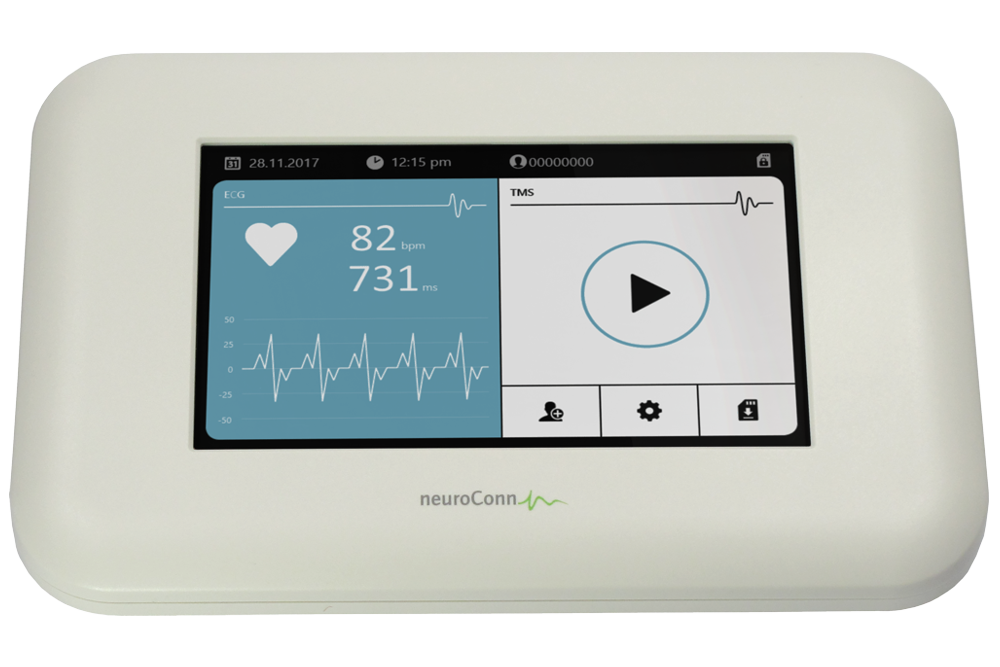
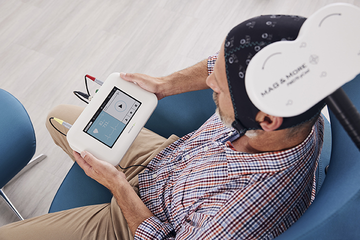
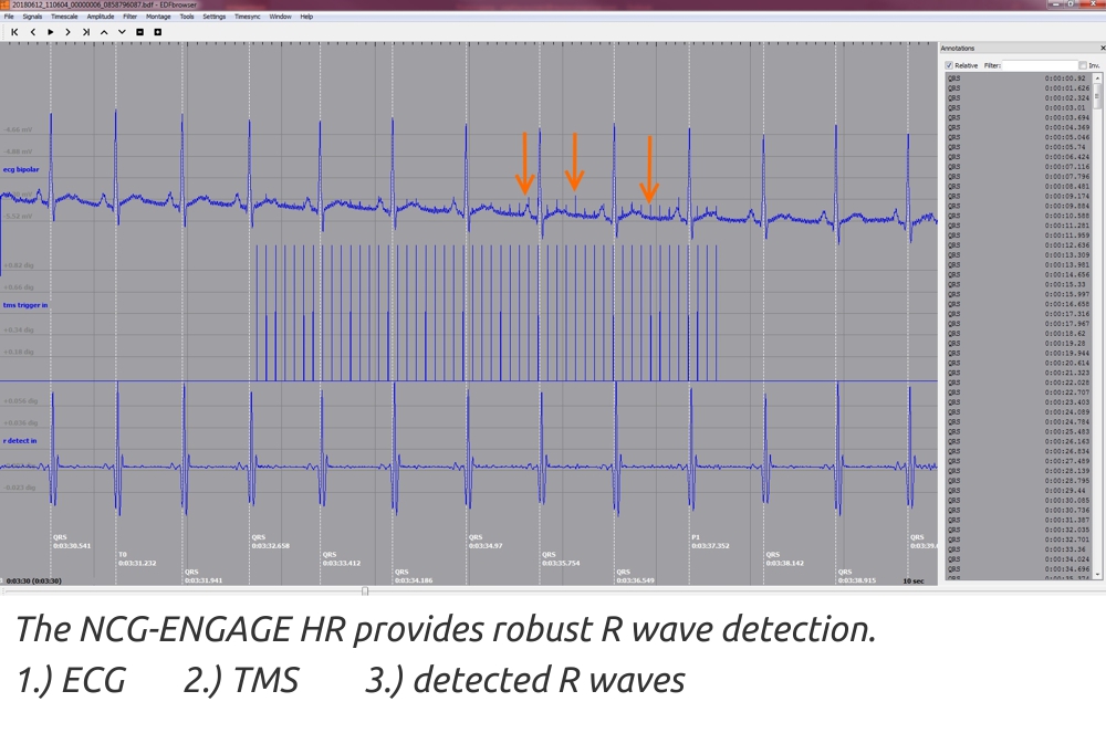
NCG-ENGAGE HR is a biofeedback device which allows the therapist to measure the heart rate during TMS of the DLPFC (dorsolateral prefrontal cortex). The autonomically working NCG-ENGAGE HR device reports analyzed heart beat intervals permanently.
.jpg)
Latest Research on Neuro-Cardiac Guided rTMS
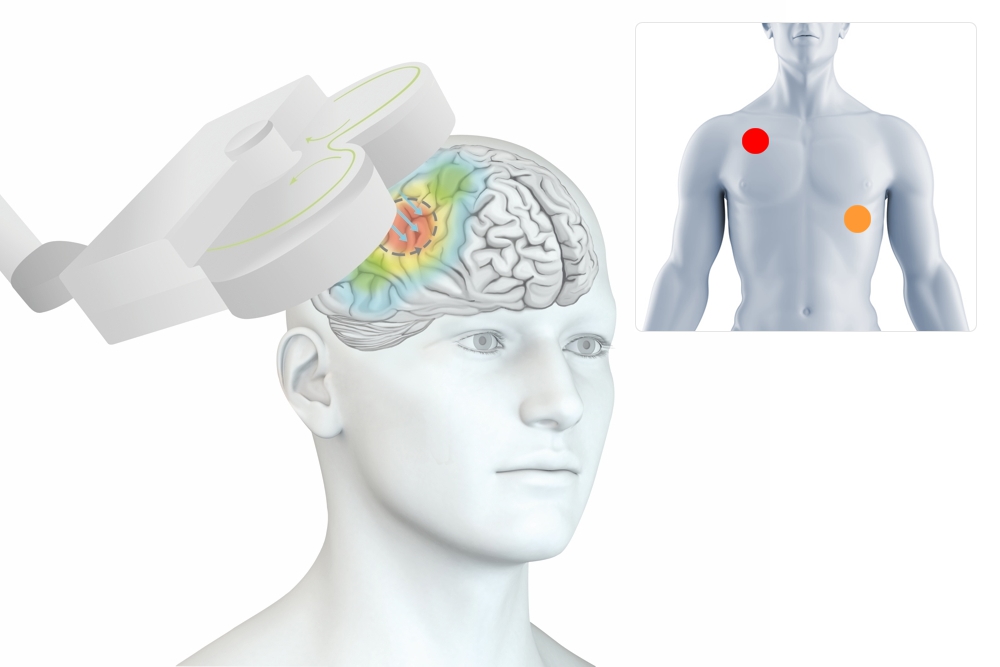
Many studies suggest the role of the connectivity of the dorsolateral prefrontal cortex (DLPFC) and the subgenual anterior cingulate cortex (sgACC) in depression. Prior research has demonstrated that neuromodulation of either of these nodes modulates the parasympathetic activity and results in a heart rate deceleration.
A new method called "Neuro-Cardiac Guided rTMS (NCG-rTMS)" helps to adequately target the frontal-vagal network. Iseger et al.1,2 show that the NCG-rTMS reliably locates the point of greatest heart rate deceleration at the DLPFC. Data from other research groups could also replicate this. Due to functional differences, this point is located at different positions in the individual patients. In many people it is located in the FC3/FC4 range. In this case rTMS treatment according to the 5 cm rule is recommended. In some people, however, it is localized around F3 or F4. Then the rTMS according to the BeamF3 method will probably be more effective.
Also, during 1 Hz brain stimulation using a ring coil, HR significantly decreased during the first 3 min of the rTMS period. Resting HR, HR during rTMS, and the degree of HR reduction were not significantly associated with treatment outcome.3
The heart–brain coupling (HBC) has a strong relationship with rTMS-entrainment of heart rate during the new TMS protocol “SAINT” developed at Stanford University (90% remission within 1 week) and has significant non-parametric correlations with meaningful clinical outcomes.4
Maximal HBC at TMS sites is negatively connected to sgACC.5, 6
Further research by Wilkening & Goya-Maldonado7 from Goettingen (Germany) showed iTBS driven heart rate modulations were associated with clinical improvement in MDD after 6 weeks. Also the White Matter microstructural properties in formix & cingulum showed predictive value for heart rate modulations and ultimately therapeutic effect
Monitoring of the frontal-vagal network seems a very promising approach for future personalized targeting where neurocares ENGAGE HR might be the prerequisite for a low-cost marker for TARGET ENGAGEMENT in rTMS treatment for MDD. Even it might be possible to use the HBC approach for thresholding at DLPFC instead of the MT.
The NCG-rTMS thus offers a way to find the most effective individual access to the frontal-vagal network via feedback of the heart rate.
1 Iseger TA et al., 2017:
Neuro-Cardiac-Guided TMS (NCG-TMS): Probing DLPFC-sgACC-vagus nerve connectivity using heart rate - First results.
(https://www.brainstimjrnl.com/article/S1935-861X(17)30793-3/abstract)
2 Iseger TA et al., 2018:
Optimizing TMS Treatment for Depression Using Cardiac Response With Neuro-Cardiac-Guided-TMS (NCG TMS) (https://www.biologicalpsychiatryjournal.com/article/S0006-3223(18)30254-3/fulltext)
Iseger TA et al., 2020:
A fronto-vagal network theory for Major Depressive Disorder: Implications for optimizing neuromodulation techniques
(https://www.sciencedirect.com/science/article/pii/S1935861X19304139)
Kaur M et al., 2020
Investigating high- and low-frequency neuro-cardiac-guided TMS for probing the frontal vagal pathway
(https://www.sciencedirect.com/science/article/pii/S1935861X20300553)
3 Sheen et. al., 2022:
Heart rate change as a predictor of treatment outcome for ring-coil accelerated low frequency repetitive transcranial magnetic stimulation in major depressive disorder: An exploratory study https://doi.org/10.1016/j.jadr.2023.100518
4 John Coetzee et al., 2023:
Heart-brain coupling in a randomized controlled trial of Standford Neuromodulation Therapy: Preliminary results and future directions. https://www.brainstimjrnl.com/article/S1935-861X(23)00664-2/pdf
5 Eva S. A. Dijkstra et. al., 2024:
BBB Probing prefrontal-sgACC connectivity using TMS-induced heart–brain coupling
https://www.nature.com/articles/s44220-024-00248-8
6 Eva S. A. Dijkstra et. al., 2023:
Transcranial Magnetic Stimulation–Induced Heart-Brain Coupling: Implications for Site Selection and Frontal Thresholding—Preliminary findings https://www.bpsgos.org/article/S2667-1743(23)00003-4/fulltext
7 Wilkening & Goya-Maldonado, 2024:
White matter tracts associated with iTBS-induced heart rate deceleration treatment response in MDD, DGPPN 2023 Berlin

NCG-ENGAGE HR: related systems
Our NCG-ENGAGE HR works with every manufacturer's TMS machine - from Magstim, MagVenture or Deymed.
We propose to use neurocare's TMS device Apollo.
